Back to Top
  • Get a comfortable stay and relax your mind

  • Enjoy holidays with our world-class service

  • Enjoy a comfortable luxury stay with us.

Showing posts with label Radiology Books. Show all posts

Clinical Radiology Made Ridiculously Simple


Description:
by:  Hugue Ouellete, Patrice Tetreault
This book was written with the intention to pursue Dr. Stephen Goldberg's vision to make
learning ridiculously simple. It is designed to rapidly teach the clinically vital components
of radiology. The reader will acquire a solid approach to radiographic examinations com-
monly interpreted without radiologist assistance, and will become comfortable at recogniz-
i ng common and dangerous conditions. Although this book is intended for medical students
and interns, it is also useful for nurses, nurse practitioners and X-ray technicians.
This small text is not meant to be a synopsis of the broad field of radiology. Radiographic
examinations interpreted by medical students and interns in the emergency room or on the
ward are emphasized, while more advanced topics are ignored. For example, plain radio-
graphs of the chest, abdomen and bones are discussed. Small chapters on the intravenous
pyelogram and computed tomography (CT) of the head are also included. However, several
topics such as barium studies, ultrasonography and magnetic resonance imaging (MRI) are
underemphasized or completely ignored.
Each chapter is subdivided into Radiographic Anatomy, Approach and Specific Problem
sections. In the Radiographic Anatomy sections, key anatomical landmarks are identified
using simplified illustrations. In the Approach sections, reading of the radiographic exami-
nation is explained using analogies, illustrative cartoons and mnemonics. In the Specific
Problem sections, the radiographic findings of the most common and deadly conditions
are discussed.
Much of this book was written in the Greek islands, where "simplicity" is a way of life. We
hope that simplicity is reflected in this work, and that you will have as much fun learning
about radiology as we did writing about it.

Basic Radiology 2nd(ID:5)

Basic Radiology (LANGE Clinical Science) 


Preface

Our main goal in this book was the creation of a concise text on current radiologic imaging for medical students and residents interested in radiology. After two introductory chapters, an organ-system approach is followed. Applicable imaging techniques and their use and indications are discussed in each organ-related chapter. Question-oriented exercises targeting common diseases in each organ system are included.

The first chapter describes the various diagnostic imaging techniques that are available: conventional radiography, nuclear medicine, ultrasonography, computed tomography, and magnetic resonance imaging. In recent years, many new techniques such as CT angiography, CT colonography, MR angiography, and MR cholangiopancreatography have emerged as new generations of CT and MR equipment have been developed. The second chapter introduces the physics of radiation and its related biological effects and basic technical considerations for ultrasound and magnetic resonance imaging. The remaining chapters focus on individual organ systems, including the heart, lungs, breast, bones, joints, abdomen, urinary tract, alimentary tract, liver, biliary system, pancreas, brain, and spine. These organ-based chapters are similarly structured for consistency. Each chapter first briefly describes the relevant recent imaging developments within each organ system and then the imaging techniques that are applicable for evaluating each organ system are reviewed and the normal anatomy is illustrated. Commonly used radiologic techniques in all areas are described in the first chapter so that repetition is avoided. Then each chapter discusses the selection of appropriate techniques with an emphasis on the proper sequencing of imaging examinations. These choices are based on the clinical presentation, the need for patient preparation and potential conflicts between techniques. The final section of these chapters is the imaging exercises with questions. Each exercise consists of numerous images and specific questions focusing on common diseases or symptoms. One question per case is used in all exercises and the case and question numbers match for clarity. A short list of recent pertinent general readings and references is included at the end of each chapter.

We hope that this book will help medical students and residents not specializing in radiology to better understand and select the many imaging modalities now available for examining their patients. Our further hope is that the interactive exercises presented will familiarize our readers with the more common diseases that current radiologic imaging can most effectively evaluate.

We wish to thank Allen D. Elster, MD Director of the Division of Radiologic Sciences and Professor and Chairman of the Department of Radiology of the Wake Forest University School of Medicine, and C. Douglas Maynard, MD, now retired former Director of Division of Radiologic Sciences and Professor and Chairman of the Department of Radiology of the Wake Forest University School of Medicine, who have provided us with the supportive environment needed to complete this endeavor. This book would also not have been possible without the able support of Martin J. Wonsiewicz, Marc Strauss, Shelley Reinhardt, and their fine associates at Lange Medical Books/McGraw-Hill and Jackie Henry at Techbooks.

A Practical Approach to Transesophageal Echocardiography


A Practical Approach to Transesophageal Echocardiography
By Albert C Perrino, Scott T Reeves


  • Publisher: Lippincott Williams & Wilkins
  • Number Of Pages: 512
  • Publication Date: 2007-11-01
  • ISBN-10 / ASIN: 0781773296
  • ISBN-13 / EAN: 9780781773294


Product Description:
Now in its Second Edition, with full-color illustrations throughout, this practical manual provides a basic introduction to the "how-to's" of diagnostic and intraoperative transesophageal echocardiography. It covers all types of heart surgery in which TEE is used and addresses clinical challenges in specific settings such as the ICU and anatomic regions such as the thoracic aorta. Each chapter includes multiple-choice questions for exam preparation. The book is portable and easy to use in the operating room. This edition includes full-color echocardiograms in all chapters and colorized and revamped drawings. A new chapter covers cardiac masses and embolic sources.


Download 
 
 OR


Angiography


Stanley Baum, Michael J Pentecost, "Abrams' Angiography: Interventional Radiology"
Lippincott Williams & Wilkins | 2005 | ISBN: 0781740894 | 1344 pages | CHM | 159 MB

The Interventional Radiology volume of the landmark reference Abrams' Angiography has now been expanded and thoroughly revised to reflect dynamic advances in interventional radiology. More than 60 contributors representing a "Who's Who" of the specialty provide comprehensive, step-by-step coverage of all contemporary vascular and nonvascular interventional procedures. Major sections discuss today's equipment and describe interventions for specific disorders of each organ system, as well as for trauma, pediatric diseases, abscess drainage, and miscellaneous disorders. Fifteen new chapters cover cutting-edge innovations, including stent-grafts, radiofrequency ablation, CT angiography, MR angiography, uterine fibroid embolization, and vertebroplasty. More than 1,100 illustrations complement the text. "Doody's Core Titles 2009."

Download


OR


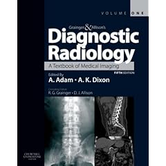
Product details

  • Hardcover: 1936 pages
  • Publisher: Churchill Livingstone; 5 edition (26 Sep 2007)
  • Language English
  • ISBN-10: 0443101639
  • ISBN-13: 978-0443101632

Product Description

Now published in 2 volumes, the new edition of this landmark text gives you a succinct but comprehensive account of all currently available imaging modalities and their clinical applications. Extensively revised and restructured, it is an excellent review text for resident radiologists, offering detailed information on all the imaging modalities and interventional procedures needed for exam preparation. Additionally, it helps the practicing radiologists with a concentrated overview of the latest imaging techniques covering all parts of the body.

Download


General Ultrasound in the Critically III by Daniel A. Lichtenstein


General Ultrasound in the Critically III by Daniel A. Lichtenstein
Publisher: Springer 2007 | ISBN: 3540736239 | PDF | 199 Pages | 14.9 MB
General ultrasound in the critically ill describes a new clinical tool: ultrasound for the intensive care and emergency physician. Written by an intensivist familiar with ultrasound, it specifically details findings of immediate clinical relevance throughout its approx. Through a whole-body approach, this book considers new emergency applications regarding the abdomen, venous system, head, heart, and the most original topic, the lung. Flow charts are proposed to resolve daily intensive care and emergency occurrences: acute dyspnea, shock, unexplained fever, etc. The strong points and pitfalls of ultrasound are reviewed in detail. This book shows just how critical ultrasound has proven to be in satisfying a major concern in the intensive care and emergency medicine fields: speed and accuracy. With this ever-present requirement for rapid diagnosis in mind, General ultrasound in the critically ill provides a key to practicing a visual medicine, a great benefit to the critically ill patient, especially since ultrasound is noninvasive and can be done at the bedside. This volume is not only an exhaustive atlas dealing with the most variable aspects of the critically ill patient, but it is above all a guide, a permanent aid in the therapeutic decision.


Download
OR

 
OR


Molecular Anatomic Imaging: PET-CT and SPECT-CT Integrated Modality Imaging, 2nd Edition

 
Gustav K von Schulthess, "Molecular Anatomic Imaging: PET-CT and SPECT-CT Integrated Modality Imaging, 2nd Edition"
Lippincott Williams & Wilkins | 2006 | ISBN: 0781776740 | 656 pages | CHM | 72,1 MB 

This fully updated Second Edition is perfect for clinicians, as it focuses sharply on clinical PET-CT and SPECT-CT examinations, omitting lengthy physics discussions. The book is now strictly disease oriented and integrates PET-CT and SPECT-CT applications completely. When both techniques are relevant for a disease, they are discussed together; when only one is relevant, it is discussed alone. More than 1,200 illustrations document the use of integrated imaging and provide very useful reference material for interpreting integrated imaging studies. A bound-in DVD contains over 80 cases to be viewed in three orthogonal planes and different CT windows organized as reference and self-assessment files. The cases provide excellent training and self-assessment material. Readers can use the DVD to enhance the book's illustrations or to test their abilities in making diagnoses on their own.


Download
 

OR 
OR 
OR

Peripheral Vascular Ultrasound

Peripheral Vascular Ultrasound
Publisher: Churchill Livingstone | ISBN: 0443072833 | edition 2005 | PDF | 244 pages | 13,2 mb
This book explores the basic scientific principles, theory, and techniques associated with peripheral vascular ultrasound and blood flow. It clearly explains how to interpret color images and Doppler spectra, as well as how to optimize scanner controls for the most effective results. Chapters include descriptions of vascular disorders, carotid duplex ultrasound, lower and upper limb arterial and venous assessment, ultrasound assessment of aneurysms, graft surveillance and pre-operative vein marking, and more. Clinical chapters also contain current criteria for grading disease. The latest technological advances, such as harmonic imaging and compound imaging, are integrated with an emphasis on safety. * Practical, step-by-step guidance on scanning shows how to perform specific procedures. * Discussions of ultrasound physics are directly tied to applications for scanning and assessing blood flow. * High-quality line drawings and images show how to perform the scan and what practitioners can expect to see. * Basic scientific principles of ultrasound instrumentation and blood flow are discussed. * Limitations and pitfalls of techniques are presented.
Download
OR

OR

Hybrid PET/CT and SPECT/CT Imaging: A Teaching File


Dominique Delbeke, Ora Israel, "Hybrid PET/CT and SPECT/CT Imaging: A Teaching File"
Springer | 2009 | ISBN: 0387928197 | 762 pages | PDF | 17 MB

Edited by Dominique Delbeke and Ora Israel, two leading authorities in the field of nuclear medicine, this practical guide is a reference source of cases for images obtained on state-of-the-art integrated PET/CT and SPECT/CT imaging systems. The cases are presented in-depth so that they will be of value to residents training in nuclear medicine and radiology and to nuclear medicine physicians and radiologists who need to become familiar with this technology. Internationally recognized contributors provide the reader with in-depth coverage on the technical and clinical aspects of hybrid imaging. Principles of hybrid imaging, physics and instrumentation, normal distribution of radiopharmaceuticals and protocols central to the field are covered. A comprehensive review of nuclear oncology cases found in everyday practice, ranging from simple to complex are also addressed. The full spectrum of clinical applications is covered including head and neck tumors, breast cancer, colorectal cancer, pancreatic cancer, and genitourinary tumors. Additional chapters examine cardiac hybrid imaging, benign bone diseases and infection and inflammation. A wealth of illustrations reinforce the key teaching points discussed throughout the book.

Download
OR

Radiology for Anaesthesia and Intensive Care


Richard Hopkins, Carol Peden, Sanjay Gandhi, "Radiology for Anaesthesia and Intensive Care"
Cambridge University Press | 2009 | ISBN: 0521735637 | 328 pages | PDF | 6,4 MB 
The advent of small, affordable ultrasound machines and the widespread use of PACS systems have made imaging more accessible to anaesthetists and intensivists than ever before. This concise, highly illustrated text discusses the key aspects of radiology, examining all imaging modalities and body regions. Introductory sections review the imaging knowledge required for the FRCA exams and the role of imaging in the Pre-Operative Assessment. These are followed by chapters on each imaging modality and body region, each containing numerous illustrations, practical advice on diagnosis, and many case illustrations. Each modality chapter contains a concise introductory section on the principles of image formation. Containing over 300 scans and illustrations, and written by a multidisciplinary team of radiologists and anaesthetists, Radiology for Anaesthesia and Intensive Care, second edition, is an invaluable aid for all anaesthetists and intensivists.
• Written by both radiologists and anaesthetists; gives a multidisciplinary perspective • Focuses on the areas of immediate relevance to anaesthesia, intensive care and perioperative medicine • Highly illustrated with over 300 high-quality scans from every relevant imaging modalityContents

Contributors; Introduction; About the FRCA examination; The pre-operative assessment; 1. Imaging the chest; 2. Imaging the abdomen; 3. Trauma radiology; 4. The cervical spine; 5. CT head; 6. Anaesthesia in the radiology department. MRI and interventional radiology; 7. Ultrasound; Index.
Review

‘… it provides examination candidates with an excellent system for in
Download
OR

OR

Medical Imaging Systems Technology


Medical Imaging Systems Technology: Methods in General Anatomy by Cornelius T. Leondes
Publisher: World Scientific Publishing 2005 | Series: Medical Imaging Systems Technology, vol.3 | 408 Pages | ISBN: 981256991X | PDF | 8.5 MB

Description: This scholarly set of well-harmonized volumes provides indispensable and complete coverage of the exciting and evolving subject of medical imaging systems. Leading experts on the international scene tackle the latest cutting-edge techniques and technologies in an in-depth but eminently clear and readable approach. Complementing and intersecting one another, each volume offers a comprehensive treatment of substantive importance to the subject areas. The chapters, in turn, address topics in a self-contained manner with authoritative introductions, useful summaries, and detailed reference lists. Extensively well-illustrated with figures throughout, the five volumes as a whole achieve a unique depth and breath of coverage. As a cohesive whole or independent of one another, the volumes may be acquired as a set or individually. 

Download


OR

The WHO Manual of Diagnostic Imaginge


The WHO Manual of Diagnostic Imaging
World Health Organization | ISBN: 9241546778 | 2006 | English | 152 Pages | PDF | 22MB

The present volume in the series of WHO manuals in diagnostic imaging, the Radiographic Anatomy and Interpretation of the Chest provides an exhaustive description of radiographic normal anatomy as well as the most common pathologic changes seen in the chest, focusing specifically on pulmonary and cardiac problems. The text aims to provide an aid to the interpretation of the chest radiograph (CXR). It is not a comprehensive account of all possible chest diseases but a descriptive text to help identify the way in which chest pathology is manifest and diagnosed on CXR.The initial chapters deal with interpretive skills and pattern recognition and the later chapters demonstrate specific pathologies. Backed by high-quality reproduction of radiographs, this manual will prove essential reading to general practitioners, medical specialists, radiographers, and radiologists in any medical settings, although focusing specifically on needs in small and mid-size hospitals.

Download


Fundamentals of Medical Imaging


Paul Suetens "Fundamentals of Medical Imaging"
Cambridge University Press | English | 2009-08-31 | ISBN: 0521519152 | 264 pages | PDF | 15 MB


Fundamentals of Medical Imaging, second edition, is an invaluable technical introduction to each imaging modality, explaining the mathematical and physical principles and giving a clear understanding of how images are obtained and interpreted. Individual chapters cover each imaging modality - radiography, CT, MRI, nuclear medicine and ultrasound - reviewing the physics of the signal and its interaction with tissue, the image formation or reconstruction process, a discussion of image quality and equipment, clinical applications and biological effects and safety issues. Subsequent chapters review image analysis and visualization for diagnosis, treatment and surgery. New to this edition: • Appendix of questions and answers • New chapter on 3D image visualization • Advanced mathematical formulae in separate text boxes • Ancillary website containing 3D animations: www.cambridge.org/suetens • Full colour illustrations throughout Engineers, clinicians, mathematicians and physicists will find this an invaluable aid in understanding the physical principles of imaging and their clinical applications.


Download
OR